
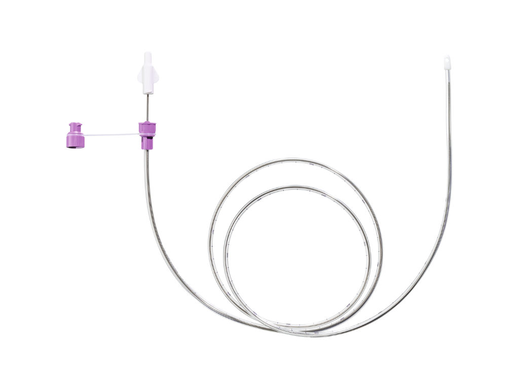
Clinical Problem
Current feeding tube verification methods are either unreliable or too time-consuming for routine use. We’re building a solution to do what current methods can’t — delivering fast, easy, and highly accurate placement verification every time.
Current Landscape
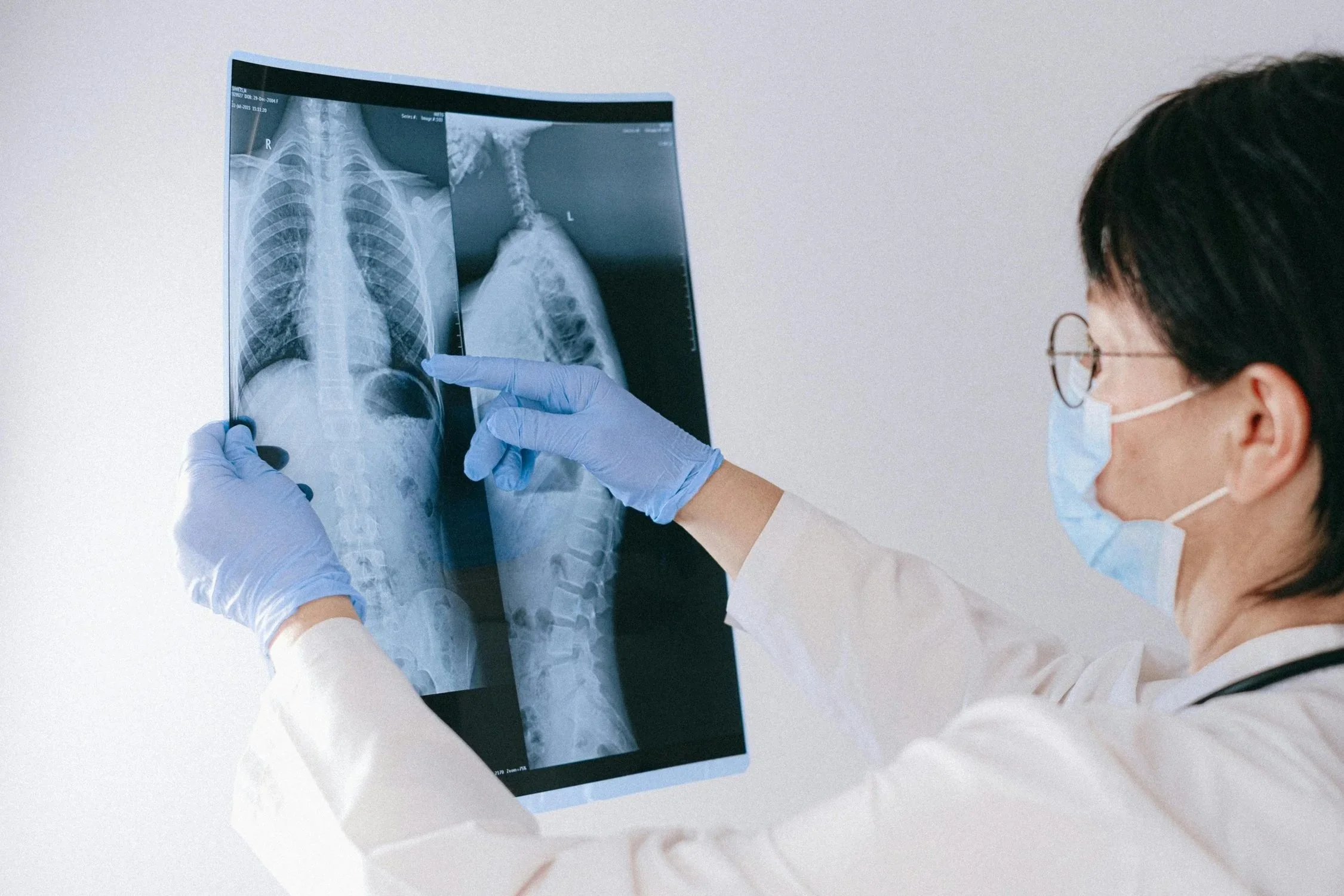
For patients who depend on feeding tubes, correct placement is a matter of safety and survival. Each year, thousands of patients experience complications from misplaced tubes, ranging from delayed nutrition to severe injury. Nurses and care teams face the difficult balance of working efficiently while ensuring absolute accuracy. The current gold standard for verification is chest x-ray, which is often time-consuming, introducing delays in care and increasing the workload for already stretched clinical staff. At its core, this challenge is about giving every patient access to safe, accurate, and timely nutrition delivery.
Challenges with X-Rays
-
Delays
X-ray verification can significantly slow down care. The feeding process must be halted until the there is time for the x-ray images to be taken, reviewed, and confirmed, often resulting in waiting hours to get results. These delays can postpone essential nutrition and medication delivery, especially for patients who rely on feeding tubes as their primary source of sustenance.
-
Cost
Each x-ray taken adds to the overall cost of care, both for hospitals and patients. Not only is the cost of the imaging factored in, but the cost of the radiologist’s expertise and time also adds to it. When multiplied across thousands of procedures, these imaging expenses quickly accumulate, straining healthcare budgets and contributing to higher patient bills.
-
Workflow Interruption
Requiring an x-ray disrupts clinical workflow and increases the workload for nurses. Staff must coordinate with radiology, arrange patient transport, and wait for confirmation before resuming feeding. This back-and-forth process takes time away from direct patient care and reduces overall efficiency in busy hospital environments.
-
Radiation Exposure
Although the radiation from a single x-ray is small, repeated imaging contributes to cumulative exposure over time, especially for patients requiring frequent tube placements or verifications. Reducing unnecessary imaging not only limits radiation risk and supports safer patient care practices.

Common Bedside Methods
Although x-rays are the gold standard, they are not always used in practice. There are some common bedside methods that nurses implement to make the verification process quicker, however, they lack the same accuracy and reliability. While offering a quick fix, these methods can compromise reliability and the safety of the patient, and thus should only be used as secondary checks.
-
Nurses use a syringe to withdraw fluid from the feeding tube and check its appearance or pH level. Gastric contents usually have a low pH (acidic), which helps indicate that the tube is in the stomach. However, this method can be unreliable if the patient is on acid-suppressing medication or if fluid cannot be easily aspirated.
-
A small amount of air is injected through the tube while the nurse listens over the stomach with a stethoscope. Hearing a “whooshing” sound suggests the tube is in the stomach, but this technique is not considered accurate or reliable since similar sounds can occur even when the tube is misplaced.
-
The external length of the tube, from the nostril to the connection point, is measured and compared to the initial insertion length. Any significant change may indicate tube movement or displacement. While simple and quick, measurement alone cannot confirm whether the tube tip is correctly positioned inside the body.